
Navigating Multiple Lipomatosis Syndrome Radiology – Have you noticed multiple lumps appearing on your body? Do they seem to be increasing in number and size? If so, you may be experiencing a condition known as Multiple Lipomatosis Syndrome (MLS).
This rare disorder is characterized by the development of multiple fatty lumps, called lipomas, under the skin. While most lipomas are harmless and can be left alone, MLS can cause discomfort and affect one’s appearance.
In order to accurately diagnose and treat MLS, radiology plays a crucial role. In this article, we will explore the causes, treatments, and removal options for MLS through the lens of Multiple Lipomatosis Syndrome Radiology.
What Are Lipomas?
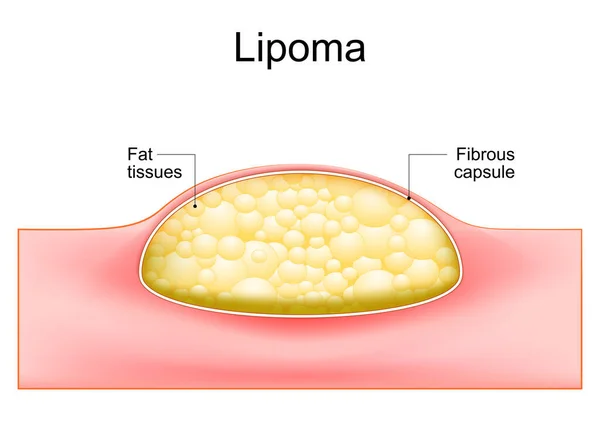
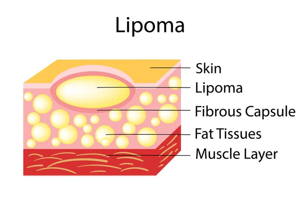
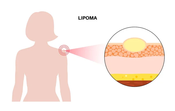
A lipoma is a growth of fat cells that form a nodule beneath the skin. Because a lipoma originates in the fatty tissue that lies between your muscle and skin, they can develop almost anywhere in the body where there is fat. They most commonly occur on the shoulders, arms, upper back, or thighs. They are typically soft, mobile, painless, and slow-growing. They usually start small and can enlarge slowly with time. Lipomas are typically benign, which means they can often be left untreated if they do not cause any issues. In rare cases, lipomas can grow and become more noticeable or uncomfortable, which should prompt evaluation by a healthcare professional.
Understanding Multiple Lipomatosis Syndrome: A Basic Overview
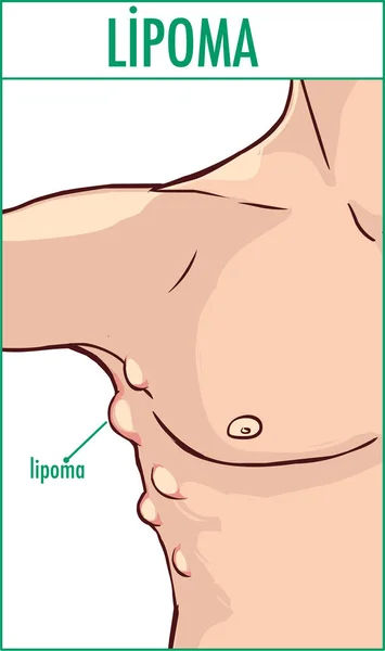
Multiple Lipomatosis Syndrome (MLS) is a rare condition that affects the development of lipomas, which are multiple fatty lumps that form beneath the skin. These lipomas can appear in various parts of the body and can range in size and number. MLS can be a cause of concern for those who experience it, as it can not only lead to discomfort but also affect one’s appearance.
Multiple lipomas typically occur in adulthood, often appearing between the ages of 30 and 60. While the exact cause of MLS is still not fully understood, there are some factors that have been associated with its development. Genetic predisposition seems to play a role, as there have been instances where MLS runs in families. Additionally, hormonal imbalances and metabolic disorders have been linked to the development of lipomas in some cases. However, it’s important to note that not all cases of MLS can be attributed to these factors, and more research is needed to fully understand the condition.
The presence of multiple lipomas can often be visually and physically distressing for those affected by MLS. The lipomas can vary in size, with some being small and barely noticeable, while others can grow larger and become more prominent. This can lead to self-consciousness and a decrease in self-esteem for individuals with MLS. Additionally, the lipomas can cause discomfort or pain, depending on their size and location. For some, even the simplest tasks like lying down or wearing certain types of clothing can become challenging due to the presence of lipomas.
In order to accurately diagnose MLS, healthcare professionals often rely on radiology. Imaging techniques such as ultrasound, CT scans, and MRI scans can provide detailed images of the lipomas and their surrounding tissues. These images help in determining the size, number, and location of the lipomas, as well as any associated complications or abnormalities. Radiology plays a crucial role in guiding treatment decisions and monitoring the progression of lipomas over time.
Understanding MLS and its impact on individuals is an important step in effectively managing the condition. By being aware of the basic overview of MLS, we can further delve into its causes, diagnosis methods, and available treatment options. In the next sections, we will explore these aspects in more detail, shedding light on the different facets of MLS and how radiology helps in its management.
Getting to the Root: What Causes Multiple Lipomatosis Syndrome?
Multiple Lipomatosis Syndrome (MLS) is a complex condition that can leave individuals with numerous questions. One of the most pressing questions is, “What causes multiple lipomas?” Understanding the root causes of MLS can help patients and healthcare professionals alike to better manage and treat this condition.
While the exact cause of MLS is still not fully understood, researchers have identified several factors that may contribute to the development of multiple lipomas. One of these factors is a genetic predisposition. There have been instances where MLS has been found to run in families, suggesting a hereditary component. Studies have shown that certain genetic mutations may increase the risk of developing lipomas.
In addition to genetic factors, hormonal imbalances, and metabolic disorders have also been linked to the development of lipomas in some cases. Hormonal imbalances, such as those seen in conditions like hypothyroidism, may disrupt the body’s natural processes and contribute to the growth of lipomas. Similarly, metabolic disorders, such as diabetes or obesity, may create an environment conducive to lipoma formation.
It’s important to note that not all cases of MLS can be attributed to these factors, and more research is needed to fully understand the condition. Other potential causes, such as trauma or injury to the affected area, are still being investigated.
Multiple lipomas are typically noncancerous growths of fat cells. They may appear as a result of an overgrowth or abnormal growth pattern of these cells. The exact triggers for this abnormal growth are still not completely understood, but the factors mentioned earlier may play a role.
Unveiling the Truth with Radiology: How Diagnosis Occurs
When it comes to diagnosing Multiple Lipomatosis Syndrome (MLS), radiology plays a crucial role in unveiling the truth behind the condition. Through the use of advanced imaging techniques, healthcare professionals are able to accurately diagnose MLS and provide patients with the necessary information for effective treatment.
One of the main imaging techniques used in diagnosing MLS is ultrasound. This non-invasive procedure uses high-frequency sound waves to create detailed images of the lipomas and surrounding tissues. Ultrasound is particularly useful in determining the size, number, and location of the lipomas, as well as any associated complications or abnormalities. It allows healthcare professionals to visualize the lumps in real time and assess their characteristics.
Another commonly used imaging technique is the CT scan or computed tomography. CT scans provide detailed cross-sectional images of the body, allowing for a more comprehensive evaluation of the lipomas. This imaging modality can provide information about the density and composition of the lipomas, as well as their relationship to nearby structures. CT scans are particularly useful in cases where lipomas are deep within the body or close to vital organs.
MRI, or magnetic resonance imaging, is another valuable tool in diagnosing MLS. This imaging technique uses powerful magnets and radio waves to generate detailed images of the body’s internal structures. MRI scans can provide clear images of the lipomas and surrounding tissues, helping healthcare professionals to assess their size, number, and location. MRI is particularly useful in cases where there is a need for more precise characterization of the lipomas or when evaluating the involvement of nearby structures.
By utilizing these advanced radiological imaging techniques, healthcare professionals can accurately diagnose MLS and develop a tailored treatment plan for each individual. The detailed images provided by radiology allow for a comprehensive evaluation of the lipomas, ensuring that no abnormalities or complications are overlooked.
Tackling the Lumps: Different Treatment Options Available
Living with Multiple Lipomatosis Syndrome (MLS) can be challenging, especially when it comes to managing the multiple fatty lumps, or lipomas, that develop under the skin. While some lipomas may be harmless and can be left alone, others may cause discomfort and affect one’s appearance. If you are dealing with MLS and want to find effective ways to tackle the lumps, there are various treatment options available to explore.
One treatment option for MLS is conservative management. This approach involves monitoring the lipomas without taking any active intervention. If the lipomas are small and do not cause any symptoms or cosmetic concerns, healthcare professionals may advise against treatment. However, regular monitoring is essential to ensure that the lipomas are not growing in size or causing any complications.
In cases where the lipomas are causing discomfort or affecting one’s quality of life, surgical intervention may be recommended. There are several surgical techniques available for lipoma removal, including excision and liposuction. Excision involves making an incision and removing the lipoma along with the surrounding tissue, while liposuction uses a suction device to remove the fatty tissue. The choice of surgical technique depends on factors such as the size and location of the lipomas, as well as individual patient preferences.
In recent years, there have also been advancements in non-surgical treatment options for MLS. One such option is injection therapy, which involves injecting medication directly into the lipoma to reduce its size or dissolve it. This technique, known as lipolysis or lipo-dissolution, can be a less invasive alternative to surgery and may be suitable for certain individuals with MLS.
It’s important to note that the effectiveness of different treatment options can vary from person to person. Factors such as the size, number, and location of the lipomas, as well as individual preferences and overall health, will be taken into consideration when determining the most appropriate treatment approach.
When Prevention Fails: Exploring Removal Procedures for Lipomas
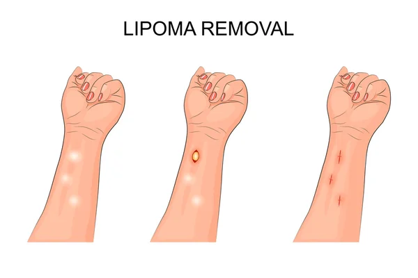
When lipomas become problematic and conservative management fails to provide relief, it may be necessary to explore removal procedures for lipomas. Surgical intervention is often the recommended course of action in these cases.
One common method of lipoma removal is excision. This involves making a small incision in the skin and removing the lipoma along with any surrounding tissue. Excision is typically reserved for larger lipomas or those located in sensitive areas. The procedure is generally performed under local anesthesia, and the incision is closed with sutures.
Liposuction is another option for lipoma removal. This procedure involves inserting a thin, hollow tube called a cannula into the skin and suctioning out the fatty tissue. Liposuction is especially effective for larger lipomas or those that are located deeper within the body. It is generally performed under local or general anesthesia, depending on the specific case.
In recent years, injection therapy has emerged as a non-surgical alternative for lipoma removal. This involves injecting medication directly into the lipoma to reduce its size or dissolve it completely. The procedure is typically performed under local anesthesia, and multiple sessions may be required to achieve the desired results.
The choice of removal procedure depends on various factors, including the size and location of the lipomas, as well as individual preferences. Your healthcare professional will evaluate your specific case and recommend the most appropriate approach.
It’s important to note that while removal procedures can effectively eliminate lipomas, there is always a risk of recurrence. Lipomas have a tendency to return, especially in individuals with Multiple Lipomatosis Syndrome. Regular monitoring and follow-up visits with your healthcare professional are crucial to detect any new or recurring lipomas.
Navigating the world of lipoma removal procedures can be overwhelming, but with the guidance of your healthcare professional, you can find the best solution for managing your lipomas. Whether you opt for surgical intervention or explore non-surgical options, the goal is to alleviate discomfort, improve your appearance, and restore your quality of life.
Life after Treatment: Managing and Monitoring Post-Procedural Outcomes
After undergoing treatment for Multiple Lipomatosis Syndrome (MLS), it is important to understand how to manage and monitor post-procedural outcomes. While treatment options can effectively eliminate lipomas, there is always a risk of recurrence, especially in individuals with MLS. To ensure the best possible outcome, it is crucial to follow certain steps in the post-treatment phase.
First and foremost, regular follow-up visits with your healthcare professional are essential. These visits allow for monitoring and early detection of any new or recurring lipomas. Your healthcare professional will assess your healing progress, check for any signs of complications, and evaluate the effectiveness of the treatment. During these visits, you will have the opportunity to discuss any concerns or questions you may have, ensuring that you are on the right track to recovery.
In addition to follow-up visits, it is important to practice good self-care and monitor your own body. Pay attention to any changes in the treated area or the appearance of new lumps. Be proactive in observing and reporting any symptoms or abnormalities to your healthcare professional. By staying vigilant and aware, you can catch any potential issues early and seek appropriate medical attention.
Managing post-procedural outcomes also involves maintaining a healthy lifestyle. This includes maintaining a balanced diet, exercising regularly, and managing any underlying health conditions. Taking care of your overall health can help minimize the risk of lipoma recurrence and promote healing.
Finally Note!
It is crucial to prioritize self-care and emotional well-being. Living with MLS and undergoing treatment can be challenging, both physically and emotionally. Take the time to practice self-care activities that promote relaxation and reduce stress. Engage in activities that boost your self-esteem and help you feel confident in your appearance.
Remember, MLS is a chronic condition, and managing post-treatment outcomes is an ongoing process. By staying proactive, maintaining regular follow-up visits, practicing good self-care, and prioritizing your emotional well-being, you can effectively manage post-procedural outcomes and live a fulfilling life after treatment. With the support of your healthcare professional and a positive mindset, you can overcome the challenges associated with MLS and enjoy improved quality of life.
Note: This article is written based on scientific evidence found by the 247newsroundtheworld.com team. Sources are duly referenced with keywords hyperlinked to source websites and are clickable for reference.
Last Updated on January 29, 2024 by 247 News Around The World